



“It’s a bit like the question of the chicken and the egg; which comes first – changes in the blood vessels in the lung, or the formation of scarring in the lung?” says CPFF Fellow Dr. Albina Tyker, now at the mid-point in her research project at McMaster University’s Firestone Institute for Respiratory Health (FIRH).
It was previously thought that scarring in areas of the lung caused abnormalities in nearby blood vessels. Recent studies, however, have shown that the relationship is more complex.
A Two-Phase Approach to Understanding ILD Progression

Dr. Tyker’s research is looking at where and how abnormal blood vessels form and most importantly, test whether the presence of abnormal blood vessels seen on routine imaging at the time of diagnosis can predict interstitial lung disease (ILD) progression over time.
There are two “phases” to her study: one that involves lab work, delving into the molecular biology of the lungs and another that analyzes CT scan results. By March 2025, Dr. Tyker had “stained” samples from 80 lung biopsies and analyzed them under a microscope. The samples came from patients with different types of ILD, including: idiopathic pulmonary fibrosis (IPF), connective tissue disease related ILD (CTD-ILD), and hypersensitivity pneumonitis (HP).
Early Findings Offer Clues About Disease Onset

Her early findings showed that blood vessels cluster around scarred areas in the lung, but are absent within the actual scar. She also found a type of collagen in small blood vessels, which is not normally found in these areas.
“The presence of this type of collagen in the walls of small blood vessels from biopsies taken from patients at the time of diagnosis suggests changes to blood vessels may take place quite early in the disease process,” says Dr. Tyker. “It indicates we are looking in the right places.”
Further analysis may help us understand how the various cells in the blood vessels and surrounding tissues are communicating and what they are telling one another to do. For instance, could signals from blood vessels be telling nearby cells to form scar tissue, she says.
Phase Two: CT Imaging for Deeper Insight
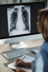
When Dr. Tyker returns to McMaster from maternity leave in September 2025, she’ll dive into the second phase of her research, looking for evidence of blood vessel abnormalities and scarring on CT scans of the lungs.
The combination of molecular data from the biopsies and data from the CT scans may provide us with new information into the processes around pulmonary fibrosis, says Dr. Tyker. And new knowledge in this area could also provide an impetus for further research into new and improved treatments.
Looking to the Future of PF Diagnosis and Treatment

Depending on the results of her research, Dr. Tyker hopes in the future it may be possible to determine the rate at which a patient can expect their disease to progress at diagnosis with a CT scan.
Dr. Tyker plans to present her findings to date at the American Thoracic Society’s conference in May 2026. She plans to apply for a second-year CPFF fellowship, to complete her research and a manuscript for publication.
Dedicated to Patients and Driven by Gratitude

“I cannot overstate how important the CPFF fellowships are to us,” says Dr. Tyker. “There is no other source of support specifically to train ILD clinician researchers in Canada. The work I’m doing now just would not be possible. I’m very grateful.”
Dr. Tyker also appreciates the CPFF resources that are available for her patients. “It is a privilege to care for such an involved and wonderful group of patients. I plan to devote my career to their care and to continue my research towards new treatments to help them.”